
Case Report
Tracheal Adenoid Cystic Carcinoma: Case Report
Unit of Otolaryngology and Head and Neck Surgery, Guatemalan Social Security Institute, Guatemala City, Guatemala.
*Corresponding Author: Howard Rosas Solares, Unit of Otolaryngology and Head and Neck Surgery, Guatemalan Social Security Institute, Guatemala City, Guatemala.
Citation: Solares HR, Monroy MYM. (2026). Tracheal Adenoid Cystic Carcinoma: Case Report, Journal of Surgical Case Reports and Reviews, BioRes Scientia Publishers. 5(1):1-3. DOI: 10.59657/2993-1126.brs.26.034
Copyright: © 2026 Howard Rosas Solares, this is an open-access article distributed under the terms of the Creative Commons Attribution License, which permits unrestricted use, distribution, and reproduction in any medium, provided the original author and source are credited.
Received: January 08, 2026 | Accepted: June 29, 2026 | Published: July 08, 2026
Abstract
Adenoid cystic carcinoma is one of the most common primary malignant tumors of the trachea, although its incidence is only 0.2 per 100,000 inhabitants. It is characterized by polypoid lesions or infiltrative plaques. It generally affects the proximal third of the trachea. The diagnosis is based on bronchoscopy to assess resectability and to take biopsies. The treatment of choice is surgical resection, and in cases with positive margins, radiotherapy. We present the case of a patient with this tumor.
Keywords: adenoid cystic carcinoma; trachea; surgery
Clinical Case Presentation
33-year-old man, with no pathological history, who presented with dyspnea and cough of 10 months' evolution. A pulmonologist ordered a computed axial tomography (CAT) scan of the neck, showing a 2 cm subglottic tumor that obstructed the airway by 50% and invaded soft tissues.
On physical examination, no lesions were observed on the nose, mouth, or pharynx. Neck without palpable lymphadenopathy. Studies were completed with a plain neck X-ray, showing almost complete obstruction of the subglottic airway by a 3 cm mass (Figure 1). A new neck CT scan showed a subglottic mass at the level of the first to fourth tracheal rings that obstructed 80% of the lumen and invaded peritracheal tissues (Figure 2). Nasolaryngoscopy was performed, observing a mass at the level of the first and second tracheal rings, with irregular edges, compromising 80% of the tracheal lumen (Figure 3). A protective tracheostomy and biopsies were performed. The histopathological result was adenoid cystic carcinoma.
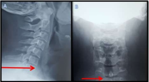
Figure 1: Image A: Anteroposterior projection of the neck x-ray. Image B: Lateral projection showing obstruction of the air column at the level of the sixth cervical vertebra in the intraluminal trachea with airway obstruction in 80% indicated with red arrow.
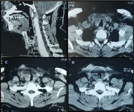
Figure 2: Computed tomography scan of the neck. Image A: A coronal projection evidencing the extension of the lesion from the subglottic region (fifth cervical vertebra), a circular mass obstructing 80% of the tracheal lumen. Image B: Axial slice at the cervical level showing a 2 cm thick circular tumor, obstructing 80% of the lumen with invasion of the soft tissues of the vocal cords, a 1 cm mass at the base of the left vocal cord. Image C: Axial slice at the level of the sixth cervical vertebra. Image D: Axial slice at the level of the seventh cervical vertebra.
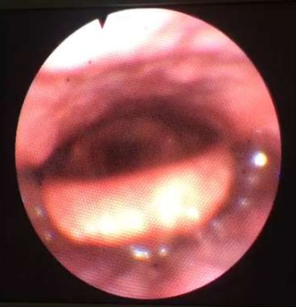
Figure 3: Flexible nasolaryngoscopy performed under sedation showing a tumor with irregular borders and obstruction of the lumen in 80%.
Resection surgery of 4 tracheal rings plus end-to-end anastomosis was performed (Figure 4A). An 8F tracheostomy cannula was left in the midline for protection of the anastomosis (Figure 4B). The histopathological diagnosis was adenoid cystic carcinoma measuring 4.5 x 4 x 3.5 cm with vascular permeation and perineural invasion infiltrating peritracheal soft tissues and thyroid on both sides; proximal, distal, and circumferential surgical margins had lesions. With this result, the patient received 35 doses of radiotherapy and 2 doses of cisplatin. Follow-up was conducted through thoracic surgery, medical oncology, and otolaryngology consultations over 5 years, using CT scans and flexible nasolaryngoscopy, without evidence of recurrence, leading to tracheostomy closure.
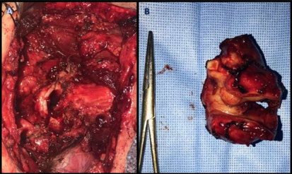
Figure 4: Figure A: The end-to-end anastomosis performed after the resection of 4 tracheal rings. Figure B: The 4.5x4x3.5 cm surgical specimen corresponding to 4 tracheal rings and the superior portion of the thyroid gland, both lobes.
Discussion
The presented case, a 33-year-old man, corresponds to an adenoid cystic carcinoma of the trachea; this pathology is more common in men, up to 70%; regarding age, the literature reports that malignant tracheal tumors are more common in people aged 60, which makes this case rare [1,3,4]. The patient, in his first evaluation, presented dyspnea with minimal effort; for this reason, the care processes were expedited, and it was decided to perform an initial intervention, which consisted of a tracheostomy to ensure ventilation, considering that, due to his age, malignant tracheal tumors are uncommon. After the report of the first biopsies, the case was reassessed to perform tumor resection surgery [3]. The largest case series includes 578 cases of malignant tracheal tumors, with a 5-year survival of 47% for localized disease, 26% for regional disease, and 4% for distant metastasis [4]. The patient described in the clinical case underwent an R1 tumor resection, so chemotherapy, radiotherapy, and close follow-up for early recurrence were considered. Several studies report that for patients with adenoid cystic carcinoma who had tumor resection and then radiotherapy, there is a 5-year recurrence rate of 52% and a 10-year recurrence rate of 29%. No studies were found that mentioned recurrence rates regarding radiotherapy and chemotherapy together as administered to the patient described in this case. However, the use of cisplatin as chemotherapy for patients with unresectable adenoid cystic carcinoma is described [3,4].
References
- Rosenberg, M., Patané, A. K., Poleri, C., Rayá, M., Rosales, A., et al. (2013). Primary Tracheal Tumors. Revista Americana de Medicina Respiratoria, 13(1):19-25.
Publisher | Google Scholor - Pearson, F. G., Todd, T. R. J., Cooper, J. D. (1984). Experience with Primary Neoplasms of The Trachea and Carina. The Journal of Thoracic and Cardiovascular Surgery, 88(4):511-518.
Publisher | Google Scholor - Rivas de Andrés, J. J., De la Torre Bravos, M. M., Blanco Gonzalez, S., Córdoba Peláez, M., Miguélez Vara, C., et al. (2000). Rare Tracheal Tumors: Adenoid Cystic Carcinoma and Mucous Gland Adenoma. Cir Esp, 67(4):391-393.
Publisher | Google Scholor - Iñiguez-García, M. A., Guzmán-de-Alba, E., Luna-Rivero, C., Salazar-Otaola, G. F., Núñez-Bustos, C. M., et al. (2014). Surgical Treatment of Adenoid Cystic Carcinoma of The Trachea: Presentation of A Case and Review of The Literature. Neumología y Cirugía de Tórax, 73(2):122-127.
Publisher | Google Scholor