
Case Report
Pseudomyxoma Peritonei: A Case Report from An Imaging Perspective
1Resident in Radiology, Department of Radiology, Camilo Cienfuegos Provincial General Hospital, Sancti Spíritus, Cuba.
2Second Degree Specialist in Family Medicine, Doctor of Medical Science, Full Professor, Associate Researcher, Directorate of Professional Training, University of Medical Sciences, Sancti Spíritus, Cuba.
3Specialist in First and Second-Degree Imaging, Master in Successful Aging, Assistant Professor, Camilo Cienfuegos Provincial General Hospital, Department of Imaging, Sancti Spíritus, Cuba.
*Corresponding Author: Belkis Alvarez Escobar, Second Degree Specialist in Family Medicine, Doctor of Medical Science, Full Professor, Associate Researcher, Directorate of Professional Training, University of Medical Sciences, Sancti Spíritus, Cuba.
Citation: Márquez EAM, Escobar BA, Pérez BMH. (2026). Pseudomyxoma Peritonei: A Case Report from An Imaging Perspective, International Clinical Case Reports and Reviews, BioRes Scientia Publishers. 4(2):1-5. DOI: 10.59657/2993-0855.brs.26.047
Copyright: © 2026 Belkis Alvarez Escobar, this is an open-access article distributed under the terms of the Creative Commons Attribution License, which permits unrestricted use, distribution, and reproduction in any medium, provided the original author and source are credited.
Received: April 17, 2026 | Accepted: July 27, 2026 | Published: August 05, 2026
Abstract
Introduction: Pseudomyxoma peritonei is a rare entity, a syndrome of progressive intraperitoneal accumulation of mucinous ascites, associated with a mucin-producing neoplasm. It has an incidence of 1 to 2 cases per million people.
Objective: To present the diagnosis of pseudomyxoma peritonei based on imaging studies.
Case Presentation: We report the case of a 65-year-old male patient with a history of type II diabetes mellitus. He presented with abdominal distension, asthenia, and lower abdominal pain. He was admitted to the General Surgery service for evaluation and treatment. Imaging studies, including abdominal ultrasound, revealed a mass occupying a large portion of the abdominal cavity, with displacement of intra-abdominal organs. The mass appeared heterogeneous, predominantly hypoechoic, with thick septa and poorly defined borders. Intra-abdominal radiopacity with displacement of intestinal loops was observed on plain abdominal x-ray. Computed tomography showed hypodensity with displacement of intestinal loops. Mesenteric cysts and very dense ascitic fluid were suspected. An exploratory laparotomy was performed, revealing an intraoperative finding of a mucinous appendiceal mass with an irregular surface and intact capsule. Post-surgical follow-up was performed using imaging.
Conclusion: Pseudomyxoma peritonei remains a challenging disease to diagnose using imaging alone. This case highlights the importance of imaging-based diagnosis and timely management. It provides a diagnostic approach using imaging that can facilitate the reporting of these rare tumors.
Keywords: pseudomyxoma peritonei; entity; imaging study; diagnostic imaging
Introduction
Pseudomyxoma peritonei is a rare condition characterized by the progressive intraperitoneal accumulation of mucinous ascites associated with a mucin-producing neoplasm [1]. It was first described by Werth in 1884, who investigated the relationship between ascites in the abdominal cavity and mucinous ovarian tumors. Later, in 1901, Fankel identified the same pathology, this time associated with mucinous appendiceal tumors. Its spread occurs through a redistribution phenomenon resulting from the free movement of epithelial cells within the peritoneal fluid and the influence of gravity [2]. Research by Zuluaga et al. [3], describes an incidence of 1 to 2 cases per million people, typically diagnosed between the ages of 40 and 55, with a higher incidence in women [4]. Regarding its etiopathogenesis, it frequently manifests as an apparently benign or well-differentiated primary tumor. Rupture of the primary tumor occurs due to the progressive secretion of mucin, and intraperitoneal dissemination of mucin takes place, where the mucin deposits follow the flow routes of the peritoneal fluid, which is conditioned by the force of gravity, directing it downwards towards the pelvis [5].
The diagnosis can be histological, with biopsy obtained through exploratory laparotomy becoming the gold standard. Recent studies have shown that ultrasound-guided fine-needle aspiration biopsy has a 95.3% success rate, even surpassing laparotomy at 93.1% [6]. Imaging, specifically ultrasound, is important in this disease. It allows visualization of collections with echogenic content, mucinous masses that may be hyperechoic or hypoechoic, and, with color Doppler, the presence or absence of vascularization. Hyperechoic foci are differentiated from other types of ascites by their mobility, as are the scalloped borders of solid organs such as the liver and spleen. Computed tomography in diagnostic imaging allows differentiation of mucinous ascites from other types of ascites, visualization of omental hyperdensity secondary to fibrosis or infiltration, and medialized intestinal loops [7]. In the province of Sancti Spíritus, at the Provincial General Hospital, there are few reports of cases diagnosed with Pseudomyxoma Peritonei. Imaging studies are essential for its diagnosis, particularly ultrasound and computed tomography (CT). Treatment is always surgical, as the condition poses a risk to the patient's life. To expand the available knowledge of this disease and aid in its imaging diagnosis, we report the case of a patient and review the literature available in the databases. The objective of this study is to present the diagnosis of Pseudomyxoma Peritonei based on imaging studies. Informed consent was obtained from the patient.
Clinical Case Presentation
A 65-year-old male patient with a history of type II diabetes mellitus reported having had a dengue arboviral infection approximately three months prior. Upon returning from a trip, he was taken to the Sancti Spíritus Provincial General Hospital due to abdominal distension, asthenia, lower abdominal pain, and scrotal and lower extremity edema. During questioning, he reported no new signs or symptoms, and was therefore admitted to the General Surgery service for further evaluation and treatment.
An abdominal ultrasound revealed a mass occupying a large portion of the abdominal cavity, with displacement of intra-abdominal organs. The mass appeared heterogeneous, predominantly hypoechoic, with thick septa, poorly defined borders, and areas of necrosis, but no calcifications were observed (Figure 1).
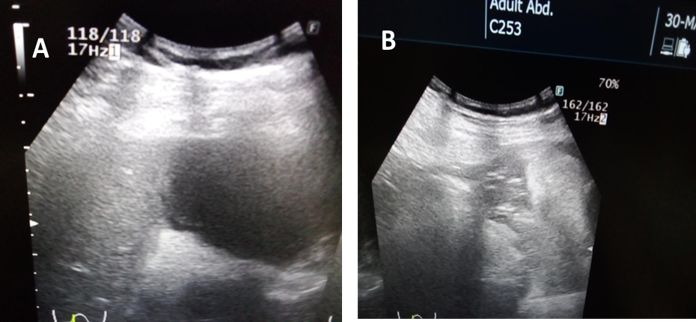
Figure 1: Echographia abdominal.
A plain abdominal x-ray reveals significant intra-abdominal opacity with displacement of intestinal loops towards the left hypochondrium and flank (Figure 2).
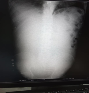
Figure 2: Plain abdominal x-ray.
The non-contrast and contrast-enhanced computed tomography (CT) scans revealed a large hypodensity occupying 80% to 90% of the abdominal cavity, with a density of 24 Hounsfield Units (HU), displacing the intestinal loops. Mesenteric cysts and very dense ascitic fluid were suspected. Periaortic lymphadenopathy was also present. A narrowing of the L5-S1 intervertebral space and diffuse changes in the trabecular bone of the right iliac bone were observed (Figure 3).
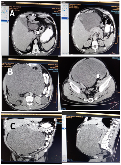
Figure 3: Contrast-enhanced CT scan of the abdomen and pelvis.
A biopsy was performed under ultrasound guidance, which did not provide elements for cytological and histological study (blood and connective tissue). Therefore, the decision was made to perform an exploratory laparotomy as the therapeutic approach (Figure 4).
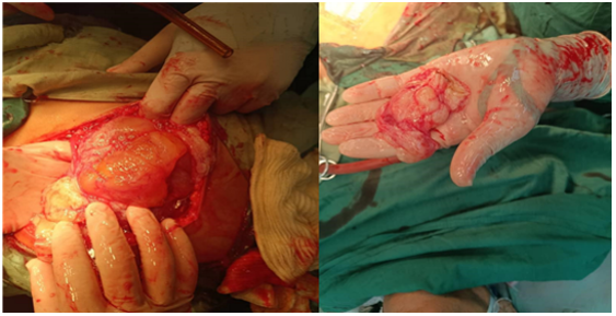
Figure 4: Exploratory laparotomy (gelatinous ascites).
The intraoperative finding during the exploratory laparotomy was a mucinous appendiceal mass with an irregular surface and intact capsule. It was accompanied by an affected peritoneum with multiple mucous formations. After the surgical intervention, the patient remained under medical and imaging follow-up.
Discussion
Pseudomyxoma peritonei is extremely rare. Its salient feature is mucinous ascites with peritoneal implants related to the rupture and dissemination of the contents of a mucinous tumor [8]. Historically, the ovary was thought to be the most frequent origin of this disease; however, more recent studies demonstrate that the most frequent primary tumor is the appendix, as in the case of Smeenk et al. [9], accounting for 82 % of cases. Other researchers suggest that appendix involvement is of metastatic origin [10,11]. The pathophysiology of the disease is determined by the accumulation, subsequent perforation, and release into the abdominal cavity of mucin produced by a tumor. This mucin disperses according to the severity of the tumor and its physiological reabsorption in certain areas of the abdomen [12]. The classification of pseudomyxoma peritonei has traditionally been confusing, leading to overlapping concepts and difficulties in comparing the efficacy of treatments. An international consensus was published that differentiated peritoneal mucinous carcinoma into low-grade, high-grade, or high-grade with signet ring cells [13]. Pseudomyxoma peritonei is more frequent in women and is diagnosed at earlier stages [14]. Although cases have been reported in men. Luque et al. [15], discuss the fundamental role of complementary imaging tests in the diagnosis of pseudomyxoma peritonei. Regarding diagnostic tests, abdominal ultrasound, in the aforementioned study, revealed ascites with septa and echoes, as well as parenchymal nodules and peritoneal masses, with hypoechoic areas in a thickened peritoneum.
In the case presented, the abdominal ultrasound imaging study revealed findings that point to the diagnosis of this entity, as did the plain abdominal x-ray, which, from an imaging perspective, provided elements for the diagnosis and management of the case. These findings coincide with the patterns identified by Ionescu et al. [16], Regarding CT, it is the imaging modality of choice, as it allows for diagnosis and assessment for subsequent medical and surgical management. Almeida et al. [17], in their case presentation, revealed a large volume of diffusely distributed intra-abdominal fluid, with an apparent mass effect in the right flank and iliac fossa, with dense intercalated septa, configuring a lobulated appearance, possibly corresponding to a cystic tumor associated with ascites or pseudomyxoma peritonei. In this case, the CT provided images that allowed for the imaging diagnosis and the decision regarding surgical treatment. Sánchez et al. [18], observed an ovoid, heterogeneous lesion with hypodense and hyperdense areas, and a scalloped appearance with the spleen and perihepatic free fluid. The present clinical case does not show hyperdense areas or calcifications in the CT scan images, unlike the one observed by the researcher.
Conclusion
Pseudomyxoma peritonei is a rare disease characterized by insidious growth. Currently, diagnosing it using imaging alone remains a challenge. This case highlights the importance of imaging-based diagnosis and timely management. Providing a diagnostic approach for pseudomyxoma peritonei through imaging can facilitate the timely diagnosis and recording of these uncommon tumors.
Declarations
Conflicts of Interest
The authors declare no conflicts of interest.
Authorship Statement
Ezequiel Alejandro Morgado: Conceptualization, data curation, formal analysis, research, methodology, supervision, visualization, project management, original draft, revised version.
Belkis Alvarez Escobar: Conceptualization, data curation, formal analysis, research, methodology, supervision. visualization. original draft. revised version.
Belkis Miladys Herrera Pérez: Conceptualization, formal analysis, research, methodology, supervision, visualization, original draft, revised version.
References
- Ning, S., Yang, Y., Wang, C., Luo, F. (2019). Pseudomyxoma Peritonei Induced by Low-Grade Appendiceal Mucinous Neoplasm Accompanied by Rectal Cancer: A Case Report and Literature Review. BMC Surgery, 19(1):42.
Publisher | Google Scholor - Morera-Ocon, F. J., Navarro-Campoy, C. (2019). History of Pseudomyxoma Peritonei from Its Origin to The First Decades of The Twenty-First Century. World Journal of Gastrointestinal Surgery, 11(9):358.
Publisher | Google Scholor - Santamaría, A. Z., Sarmiento Serrano, J. R., Cock Botero, A. M., Uribe González, R., Osorio Castrillón, L. M., et al. (2015). Neoplasias Mucinosas del Apéndice. Rev Colomb Radiol, 4252-4259.
Publisher | Google Scholor - Yu, B., Raj, M. S. (2024). Pseudomyxoma Peritonei. StatPearls [Internet].
Publisher | Google Scholor - Gómez, C. H., Bustillos, R. C., Meliá, B. W., García, M. Á., Garijo, T. G., et al. (2024). Revisión del Pseudomixoma Peritoneal, Una Patología Infrecuente. Seram, 1(1).
Publisher | Google Scholor - Corral-Vázquez, R. A. D., Tafoya Hernández, J. O., Parra Macías, A. (2024). Pseudomixoma Peritoneal: Reporte de Un Caso. Revista de la Facultad de Medicina, 67(3):32-36.
Publisher | Google Scholor - Kopereck, V., Mezzomo, B., Camillo, R., Camillo, B. B., Teixeira, C. M. (2024). Pseudomixoma Peritoneal: Um Diagnóstico de Difícil Realização. Revista Foco, 17(3):e4768-e4768.
Publisher | Google Scholor - Mittal, R., Chandramohan, A., Moran, B. (2017). Pseudomyxoma Peritonei: Natural History and Treatment. International Journal of Hyperthermia, 33(5):511-519.
Publisher | Google Scholor - Smeenk, R. M., Van Velthuysen, M. L. F., Verwaal, V. J., Zoetmulder, F. A. N. (2008). Appendiceal Neoplasms and Pseudomyxoma Peritonei: A Population-Based Study. European Journal of Surgical Oncology, 34(2):196-201.
Publisher | Google Scholor - Muñoz, E., Marco, C., Veloso, E., Bretcha, P., Rodríguez Santiago, J., et al. (2000). Seudomixoma Peritoneal de Origen Apendicular. Cir. Esp, 464-466.
Publisher | Google Scholor - Govaerts, K., Lurvink, R. J., De Hingh, I. H. J. T., Van der Speeten, K., Villeneuve, L., et al. (2021). Appendiceal Tumours and Pseudomyxoma Peritonei: Literature Review With PSOGI/EURACAN Clinical Practice Guidelines for Diagnosis and Treatment. European Journal of Surgical Oncology, 47(1):11-35.
Publisher | Google Scholor - Bignell, M., Carr, N. J., Mohamed, F. (2016). Pathophysiology and Classification of Pseudomyxoma Peritonei. Pleura and Peritoneum, 1(1):3-13.
Publisher | Google Scholor - Carr, N. J., Cecil, T. D., Mohamed, F., Sobin, L. H., Sugarbaker, P. H., et al. (2016). A Consensus for Classification and Pathologic Reporting of Pseudomyxoma Peritonei and Associated Appendiceal Neoplasia: The Results of The Peritoneal Surface Oncology Group International (PSOGI) Modified Delphi Process. The American Journal of Surgical Pathology, 40(1):14-26.
Publisher | Google Scholor - Gallardo-Martínez, J., Palomo-Rodríguez, M. F., Brenner-Anidjar, R. D., Márquez-Maraver, F., Pantoja-Garrido, M., et al. (2022). Pseudomixoma Peritoneal Primario de Origen Ovárico. Ginecología y Obstetricia de México, 90(8):706-712.
Publisher | Google Scholor - Luque-González, P., Sutil, L. A., Cabezas-Palacios, M. N., Jiménez-Gallardo, J., Rodríguez-Jiménez, I. (2021). Peritonei Pseudomyxoma Diagnosed in The Context of A High Suspicion of Ovarian Carcinoma: A Case Report. Ginecología y obstetricia de México, 89(7):549-555.
Publisher | Google Scholor - Ionescu, S., Marincas, M., Madge, O. L., Dicu-Andreescu, I. G., Chitoran, E., et al. (2024). Ovarian Causes of Pseudomyxoma Peritonei (PMP)-A Literature Review. Cancers, 16(8):1446.
Publisher | Google Scholor - de Noronha, A. A., da Silva, W. L. D., Jomah Filho, J. B. R., da Silva, W. R. C., de Menezes, I. (2022). Pseudomixoma Peritoneal-Relato de Caso. COORTE-Revista Científica do Hospital Santa Rosa, (13):136-142.
Publisher | Google Scholor - Sánchez Silva, M. C., Morales Pérez, F. (2021). Pseudomixoma Peritoneal. Acta Médica Grupo Ángeles, 19(1):134-135.
Publisher | Google Scholor