Review Article
Artificial Intelligence in Oral Potentially Malignant Disorders: From Early Detection to Precision Risk Assessment
Department of Oral Medicine and Radiology, KAHER’s KLE Vishwanath Katti Institute of Dental Sciences, Belagavi, Karnataka, India.
*Corresponding Author: Niyati Shah, Department of Oral Medicine and Radiology, KAHER’s KLE Vishwanath Katti Institute of Dental Sciences, Belagavi, Karnataka, India.
Citation: Shah N. (2026). Artificial Intelligence in Oral Potentially Malignant Disorders: From Early Detection to Precision Risk Assessment. Dentistry and Oral Health Care, BioRes Scientia Publishers. 5(2):1-6. DOI: 10.59657/2993-0863.brs.26.064
Copyright: © 2026 Niyati Shah, this is an open-access article distributed under the terms of the Creative Commons Attribution License, which permits unrestricted use, distribution, and reproduction in any medium, provided the original author and source are credited.
Received: July 20, 2026 | Accepted: August 10, 2026 | Published: August 21, 2026
Abstract
Artificial intelligence (AI) has emerged as a transformative technology in healthcare, offering novel opportunities for the early detection, diagnosis, and risk assessment of Oral Potentially Malignant Disorders (OPMDs). OPMDs, including oral leukoplakia, oral erythroplakia, oral lichen planus, and oral submucous fibrosis, possess varying risks of malignant transformation into oral squamous cell carcinoma (OSCC), making timely diagnosis essential for improving patient outcomes. Recent advances in machine learning (ML), deep learning (DL), convolutional neural networks (CNNs), computer vision, and digital pathology have enabled the development of AI-assisted systems capable of analyzing clinical photographs, histopathological images, and multimodal clinical datasets with high accuracy and consistency. These technologies have demonstrated promising performance in automated lesion detection, lesion classification, epithelial dysplasia assessment, prediction of malignant transformation, and clinical decision support. Furthermore, the integration of AI with digital pathology, molecular biomarkers, and predictive analytics has advanced personalized risk stratification and precision oral oncology. Despite these promising developments, challenges related to dataset quality, algorithm transparency, external validation, ethical considerations, and regulatory approval continue to limit routine clinical implementation. This narrative review summarizes the current evidence regarding AI applications in the detection and risk assessment of OPMDs, discusses their clinical utility and existing limitations, and highlights future directions for integrating AI into precision oral healthcare. AI should be regarded as a valuable adjunct to clinical expertise, with the potential to improve early diagnosis, optimize patient management, and ultimately reduce the burden of oral cancer through timely intervention.
Keywords: artificial intelligence; oral potentially malignant disorders; oral leukoplakia; oral lichen planus; oral submucous fibrosis; oral erythroplakia; oral squamous cell carcinoma; ML; DL; digital pathology; precision oral oncology
Introduction
Oral Potentially Malignant Disorders (OPMDs) constitute a heterogeneous group of oral mucosal lesions associated with an increased risk of malignant transformation into oral squamous cell carcinoma (OSCC) [1-3]. Early detection and accurate diagnosis are essential for reducing disease progression and improving patient outcomes. However, OPMDs often exhibit diverse clinical presentations, overlapping features with benign lesions, and unpredictable biological behavior, making diagnosis and risk assessment challenging [4,5]. Conventional diagnostic approaches, including clinical examination and histopathological evaluation, remain the gold standard but are susceptible to observer variability, sampling errors, delayed diagnosis, and limited ability to predict malignant transformation [5,6].
The rapid evolution of artificial intelligence (AI) has introduced promising opportunities to address these challenges. AI refers to computational systems capable of performing tasks that traditionally require human intelligence, including pattern recognition, image analysis, decision-making, and predictive modeling [7]. Through machine learning (ML) and deep learning (DL) algorithms, AI can analyze large volumes of clinical photographs, radiographic images, histopathological slides, and molecular data with greater speed, consistency, and accuracy than conventional methods [7,8].
The integration of AI into OPMD diagnosis represents a significant advancement toward precision oral healthcare. AI-assisted technologies have demonstrated considerable potential in automated lesion detection, lesion classification, digital pathology, biomarker analysis, and prediction of malignant transformation risk, thereby supporting earlier diagnosis and more objective clinical decision-making [8-10]. This narrative review critically evaluates the current evidence on AI applications in the detection and risk assessment of Oral Potentially Malignant Disorders and discusses their clinical utility, existing limitations, and future prospects in precision oral oncology.
Fundamentals of Artificial Intelligence in Oral Mucosal Disease Diagnosis
Artificial intelligence (AI) comprises a range of computational technologies that enable machines to perform tasks requiring human intelligence, including image recognition, pattern analysis, prediction, and clinical decision support. In the field of oral mucosal diseases, particularly Oral Potentially Malignant Disorders (OPMDs), AI has emerged as a valuable tool for improving early detection, lesion classification, risk stratification, and prediction of malignant transformation. By integrating clinical images, histopathological data, molecular biomarkers, and patient information, AI-based systems can assist clinicians in making more accurate, objective, and timely diagnostic decisions [1-3].
Table 1: Core Artificial Intelligence Technologies and Their Applications in Oral Mucosal Diseases.
| Technology | Definition | Application in Oral Mucosal Diseases/OPMDs |
| Artificial Intelligence (AI) | Simulation of human intelligence using computational systems | Clinical decision support for diagnosis and management of oral mucosal lesions |
| Machine Learning (ML) | Algorithms that learn patterns from data without explicit programming | Prediction of lesion behavior and malignant transformation risk |
| Deep Learning (DL) | Advanced ML using multilayer neural networks for automated feature extraction | Automated classification of oral mucosal lesions from clinical and histopathological images |
| Convolutional Neural Networks (CNNs) | Deep learning architecture specialized for image analysis | Detection and classification of leukoplakia, erythroplakia, oral lichen planus, and other OPMDs from clinical photographs |
| Computer Vision | Automated interpretation and analysis of digital images | Segmentation, localization, and quantitative assessment of oral mucosal lesions |
| Natural Language Processing (NLP) | AI technique for understanding and processing clinical text | Extraction of diagnostic information from electronic health records and pathology reports |
| Generative AI | AI models capable of generating text, images, or clinical recommendations | Clinical documentation, differential diagnosis support, and educational assistance |
| Predictive Analytics | Statistical and AI-based models for forecasting clinical outcomes | Prediction of malignant transformation, recurrence, and patient-specific risk stratification |
Oral Potentially Malignant Disorders (OPMDs), including oral leukoplakia, oral erythroplakia, oral lichen planus, and oral submucous fibrosis, represent a group of chronic oral mucosal lesions with varying risks of progression to oral squamous cell carcinoma (OSCC). Early detection and accurate risk assessment are critical for improving prognosis and reducing oral cancer-related morbidity and mortality. However, conventional diagnosis relies primarily on clinical examination and histopathological assessment, both of which are subject to observer variability and have limited ability to predict malignant transformation [11-13].
Artificial intelligence (AI) has emerged as a promising adjunctive tool for improving the detection and characterization of OPMDs. Machine learning (ML) and deep learning (DL) algorithms can identify subtle morphological and textural changes in oral mucosal lesions that may not be readily apparent during routine clinical examination. By analyzing standardized intraoral photographs, AI systems can assist in the automated detection and classification of suspicious lesions, thereby enhancing diagnostic consistency and supporting early clinical decision-making [12,14].
Among deep learning techniques, Convolutional Neural Networks (CNNs) have demonstrated considerable success in image-based analysis of oral mucosal lesions. These models evaluate lesion characteristics such as color variation, border irregularity, surface texture, lesion size, and morphological features to differentiate benign lesions from OPMDs and to identify lesions with a higher probability of epithelial dysplasia or malignant transformation [14-16].
Beyond image interpretation, AI has expanded into multimodal risk prediction by integrating clinical findings, demographic characteristics, tobacco and areca nut exposure, histopathological features, and molecular biomarkers. Such predictive models enable individualized risk stratification, helping clinicians identify patients who require early biopsy, closer surveillance, or prompt therapeutic intervention. This personalized approach has the potential to optimize clinical management while minimizing unnecessary invasive procedures [15-18].
AI has also demonstrated significant utility in digital pathology. Deep learning algorithms can analyze whole-slide histopathological images, detect epithelial dysplasia, quantify nuclear atypia, evaluate architectural abnormalities, and identify microscopic features associated with malignant progression. These systems can improve diagnostic reproducibility, reduce interpretation time, and provide valuable decision support for oral pathologists [16-18].
Table 2: Artificial Intelligence Approaches for the Detection and Risk Assessment of Oral Potentially Malignant Disorders.
| Clinical Application | AI Technique | Parameters Measured | Clinical Outcome |
| Automated lesion Detection | Convolutional Neural Networks (CNNs) | Lesion size, shape, border irregularity, colour distribution, surface texture | Early identification of suspicious oral mucosal lesions |
| Lesion Classification | Deep Learning (DL) | Morphological and colour characteristics of oral lesions | Differentiates benign lesions, OPMDs, and oral squamous cell carcinoma |
| Epithelial Dysplasia Assessment | Machine Learning (ML) with Digital Pathology | Nuclear atypia, epithelial thickness, keratinization, mitotic figures, tissue architecture | Objective grading of epithelial dysplasia |
| Malignant Transformation Prediction | Multimodal ML models | Clinical findings, lesion characteristics, tobacco/areca nut history, histopathological features, molecular biomarkers | Individualized risk prediction and surveillance planning |
| Histopathological Image Analysis | AI-assisted Whole-Slide Imaging (WSI) | Cellular morphology, inflammatory infiltrate, nuclear-cytoplasmic ratio, tissue organization | Improved diagnostic consistency and reduced observer variability |
| Clinical Decision Support | Predictive AI Algorithms | Combined clinical, imaging, histopathological, and biomarker data | Supports biopsy decisions, treatment planning, and follow-up scheduling |
AI-based diagnosis of OPMDs begins with the acquisition of standardized intraoral photographs or digitized histopathological slides. Before analysis, images undergo preprocessing procedures such as colour normalization, contrast enhancement, noise reduction, and lesion segmentation to improve image quality and isolate the region of interest [20]. Deep learning algorithms, particularly Convolutional Neural Networks (CNNs), subsequently extract hierarchical image features without manual intervention, enabling automated recognition of lesion-specific characteristics including colour heterogeneity, border irregularity, surface texture, lesion dimensions, and morphological asymmetry [21]. Following feature extraction, the trained model compares these characteristics with patterns learned from large annotated datasets to classify lesions as benign, OPMDs, or oral squamous cell carcinoma. In advanced AI systems, image-derived information is integrated with clinical variables such as patient age, sex, tobacco and areca nut habits, lesion duration, anatomical location, histopathological findings, and molecular biomarkers. This multimodal analysis generates individualized risk scores that estimate the probability of epithelial dysplasia or malignant transformation, thereby assisting clinicians in determining the need for biopsy, treatment, and follow-up intervals [20,21].
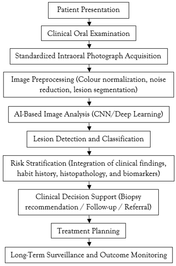
AI-Driven Clinical Workflow for the Detection and Risk Assessment of Oral Potentially Malignant Disorders (OPMDs)
Advantages of Artificial Intelligence in Oral Mucosal Disease Diagnosis
Artificial intelligence (AI) has emerged as a promising adjunct in the diagnosis and management of oral mucosal diseases, particularly Oral Potentially Malignant Disorders (OPMDs). One of its greatest advantages is its ability to improve diagnostic accuracy by objectively analyzing clinical photographs and histopathological images, thereby reducing observer variability and minimizing diagnostic errors [20,21]. AI algorithms can detect subtle alterations in lesion morphology, colour, texture, and border characteristics that may not be readily apparent during routine clinical examination, facilitating earlier recognition of high-risk lesions.
AI also enables automated lesion detection and classification, assisting clinicians in distinguishing benign oral mucosal lesions from OPMDs and oral squamous cell carcinoma (OSCC). Integration of clinical information with histopathological features, patient demographics, tobacco and areca nut exposure, and molecular biomarkers allows AI models to perform individualized risk stratification and predict the likelihood of malignant transformation [20-22]. Such predictive capabilities may improve patient selection for biopsy, optimize surveillance intervals, and support evidence-based clinical decision-making.
Another important advantage is the application of AI in digital pathology. Deep learning algorithms can evaluate whole-slide histopathological images, quantify epithelial dysplasia, detect nuclear atypia, and assess tissue architecture with high reproducibility. These systems enhance diagnostic consistency, reduce interpretation time, and provide valuable decision support for oral pathologists [21-23]. Furthermore, AI-driven analysis of large clinical datasets may facilitate the identification of novel prognostic biomarkers and improve understanding of disease progression, contributing to more personalized management strategies.
Limitations and Challenges
Despite its considerable potential, several challenges continue to limit the routine clinical implementation of AI in the diagnosis of oral mucosal diseases. High-performing AI models require large, diverse, and accurately annotated datasets; however, many published studies are based on relatively small, retrospective, or single-center datasets, limiting their generalizability across different populations [22,23]. Variations in image quality, acquisition protocols, lighting conditions, lesion appearance, and annotation methods further affect model performance and reproducibility.
The limited interpretability of many deep learning algorithms, often described as the "black-box" problem, remains another significant concern because clinicians may find it difficult to understand the reasoning behind AI-generated predictions [23]. Ethical and legal issues related to patient privacy, data security, informed consent, algorithmic bias, and medico-legal accountability must also be addressed before widespread adoption. In addition, regulatory approval, external validation, integration into existing clinical workflows, and the costs associated with digital infrastructure and workforce training remain important barriers to implementation, particularly in low-resource healthcare settings [22-24].
Future Perspectives
Future developments in AI are expected to further improve the early detection and risk assessment of OPMDs through the integration of multimodal clinical data. Combining standardized clinical photographs with histopathological findings, molecular biomarkers, genomic information, and lifestyle factors such as tobacco and areca nut exposure will enable more accurate prediction of malignant transformation and support personalized patient management [22-24].
Advances in explainable AI are anticipated to improve transparency by allowing clinicians to visualize the image features that contribute to diagnostic decisions, thereby increasing confidence in AI-assisted systems. Integration of AI with digital pathology, whole-slide imaging, and point-of-care diagnostic technologies may facilitate rapid screening, objective dysplasia grading, and real-time clinical decision support. Prospective multicenter studies, standardized image databases, and robust external validation will be essential to ensure reliable and equitable implementation of AI across diverse healthcare settings.
Conclusion
Artificial intelligence is rapidly transforming the diagnosis and management of Oral Potentially Malignant Disorders by enhancing lesion detection, improving diagnostic accuracy, supporting histopathological assessment, and enabling individualized risk prediction. Current evidence suggests that AI can serve as a valuable clinical decision-support tool for the early identification of lesions with malignant potential, thereby facilitating timely intervention and improving patient outcomes. Nevertheless, challenges related to dataset quality, algorithm transparency, regulatory approval, and ethical governance remain to be addressed before widespread clinical adoption. Continued advances in explainable AI, multimodal data integration, and prospective clinical validation are expected to accelerate the incorporation of AI into routine practice and support the development of precision oral oncology.
Conflicts of Interest
The author has no conflicts of interest to declare.
References
- Li, H., Chen, S., Chang, B., Wang, X., He, Y., et al. (2026). Application of Artificial Intelligence in Oral Health Management: Challenges and Opportunities. Frontiers in Medicine, 13:1700529.
Publisher | Google Scholor - Agrawal, P., Nikhade, P., Nikhade, P. P. (2022). Artificial Intelligence in Dentistry: Past, Present, and Future. Cureus, 14(7).
Publisher | Google Scholor - Andrzejczak, B., Diedul, A., Szczepankiewicz, A., Trojanowski, P., Skrzypczak, A., et al. (2026). Use of Artificial Intelligence for Diagnosing Oral Mucosa Conditions: A Review. Diagnostics, 16(2):365.
Publisher | Google Scholor - Ossowska, A., Kusiak, A., Świetlik, D. (2022). Artificial Intelligence in Dentistry-Narrative Review. International Journal of Environmental Research and Public Health, 19(6):3449.
Publisher | Google Scholor - Cai, X. J., Peng, C. R., Cui, Y. Y., Li, L., Huang, M. W., et al. (2025). Identification of Genomic Alteration and Prognosis Using Pathomics-Based Artificial Intelligence in Oral Leukoplakia and Head and Neck Squamous Cell Carcinoma: A Multicenter Experimental Study. International Journal of Surgery, 111(1):426-438.
Publisher | Google Scholor - Carrillo-Perez, F., Pecho, O. E., Morales, J. C., Paravina, R. D., Della Bona, A., et al. (2022). Applications of Artificial Intelligence in Dentistry: A Comprehensive Review. Journal of Esthetic and Restorative Dentistry, 34(1):259-280.
Publisher | Google Scholor - Meghil, M. M., Rajpurohit, P., Awad, M. E., McKee, J., Shahoumi, L. A., et al. (2022). Artificial Intelligence in Dentistry. Dentistry Review, 2(1):100009.
Publisher | Google Scholor - Khanna, S. S., Dhaimade, P. A. (2017). Artificial Intelligence: Transforming Dentistry Today. Indian Journal of Basic and Applied Medical Research, 6(3):161-167.
Publisher | Google Scholor - Grischke, J., Johannsmeier, L., Eich, L., Griga, L., Haddadin, S. (2020). Dentronics: Towards Robotics and Artificial Intelligence in Dentistry. Dental Materials, 36(6):765-778.
Publisher | Google Scholor - Lee, S. J., Poon, J., Jindarojanakul, A., Huang, C. C., Viera, O., et al. (2025). Artificial Intelligence in Dentistry: Exploring Emerging Applications and Future Prospects. Journal of Dentistry, 155:105648.
Publisher | Google Scholor - Nguyen, T. T., Larrivée, N., Lee, A., Bilaniuk, O., Durand, R. (2021). Use of Artificial Intelligence in Dentistry: Current Clinical Trends and Research Advances. Journal of the Canadian Dental Association, 87(l7):1488-2159.
Publisher | Google Scholor - Ahmed, N., Abbasi, M. S., Zuberi, F., Qamar, W., Halim, M. S. B., et al. (2021). Artificial Intelligence Techniques: Analysis, Application, and Outcome in Dentistry-A Systematic Review. BioMed Research International, 1:9751564.
Publisher | Google Scholor - Thurzo, A., Urbanova, W., Novak, B., Czako, L., Siebert, T., et al. (2022). Where is The Artificial Intelligence Applied in Dentistry? Systematic Review and Literature Analysis. Healthcare, 10(7):1269.
Publisher | Google Scholor - Kukreja, P. (2025). Integration of Artificial Intelligence in Dentistry: A Systematic Review of Educational and Clinical Implications. Cureus, 17(2):e79350-e79350.
Publisher | Google Scholor - Achararit, P., Manaspon, C., Jongwannasiri, C., Phattarataratip, E., Osathanon, T., et al. (2023). Artificial Intelligence-Based Diagnosis of Oral Lichen Planus Using Deep Convolutional Neural Networks. European Journal of Dentistry, 17(04):1275-1282.
Publisher | Google Scholor - Feher, B., Tussie, C., Giannobile, W. V. (2024). Applied Artificial Intelligence in Dentistry: Emerging Data Modalities and Modeling Approaches. Frontiers in Artificial Intelligence, 7:1427517.
Publisher | Google Scholor - Samaranayake, L., Tuygunov, N., Schwendicke, F., Osathanon, T., Khurshid, Z., et al. (2025). The Transformative Role of Artificial Intelligence in Dentistry: A Comprehensive Overview. Part 1: Fundamentals of AI, And Its Contemporary Applications in Dentistry. International Dental Journal, 75(2):383-396.
Publisher | Google Scholor - El Joudi, N. A., Othmani, M. B., Bourzgui, F., Mahboub, O., Lazaar, M. (2022). Review of The Role of Artificial Intelligence in Dentistry: Current Applications and Trends. Procedia Computer Science, 210:173-180.
Publisher | Google Scholor - Thurzo, A., Strunga, M., Urban, R., Surovková, J., Afrashtehfar, K. I. (2023). Impact of Artificial Intelligence on Dental Education: A Review and Guide for Curriculum Update. Education Sciences, 13(2):150.
Publisher | Google Scholor - Hao, Y., Zhou, M., Jie, W., Tang, F., Zhang, S., et al. (2023). Deep Learning Algorithms for Classification and Detection of Recurrent Aphthous Ulcerations Using Oral Clinical Photographic Images. Journal of Dental Sciences, 19(1):254-260.
Publisher | Google Scholor - Mörch, C. M., Atsu, S., Cai, W., Li, X., Madathil, S. A., et al. (2021). Artificial Intelligence and Ethics in Dentistry: A Scoping Review. Journal of Dental Research, 100(13):1452-1460.
Publisher | Google Scholor - Ghods, K., Azizi, A., Jafari, A., Ghods, K. (2023). Application of Artificial Intelligence in Clinical Dentistry, A Comprehensive Review of Literature. Journal of Dentistry, 24(4):356.
Publisher | Google Scholor - Lu, M., Zhang, J., Cao, Y., Zhang, R., Zhang, G. (2024). Diagnosing Oral Mucosal Diseases Using Deep Learning. In 2024 China Automation Congress (CAC). IEEE. 1300-1305.
Publisher | Google Scholor - Sakharkar, M., Spokas, G., Berry, L., Daniels, K., Nithagon, P., et al. (2025). Non-Invasive Screening for Laryngeal Cancer Using the Oral Cavity as A Proxy for Differentiation of Laryngeal Cancer Versus Leukoplakia: A Novel Application of ESS Technology and Artificial Intelligence Supported Statistical Modeling. American Journal of Otolaryngology, 46(1):104581.
Publisher | Google Scholor